How did the first person to see a virus know that it was a virus without any references as to what a virus looks like?
The Discovery of the Virus was done by Process of Elimination, not Direct Observation.
Two scientists D. Ivanovski and M. Beijerinck, used process of elimination to discover the first virus(es). Bacterium are a type of living organism (amongst the single celled organisms and such) known as 'contagium fixium' (in their native tongue) and in English this translates to 'infectious being fixed in place' but they discovered a new kind of infectious being; 'contagium vivum fluidum' which, in English, is an infectious soluble agent.[1] This agent, be it for instance Agar Gel, renders the bacteria still and thus immovable as they can't migrate through it as the density is too high. The density, however, was not too high for the pathogens that Ivanovski and Beijerinck tested, this led to them naming this new type a Tobacco mosaic virus (the 'virus' being the key part of it).[1]
How do viruses find their host if they have no legs, arms, eyes, ears, brains, sense of touch or means of locomotion?
Pro is conceding that they can exist, and is merely asking me to clarify some specifics here.
Question 1: What makes viruses end up infecting one type of host-cell per type of virus?
Question 2: How do they do that and even move if they have no limbs or nerve-based senses?
Before I answer both, it is important to know the steps involved in the virus-infecting process in order to 'map-out' the events in your brain.
The Lyctic cycle is the way to answer the Question 1.[2]
During the lytic cycle of virulent phage, the bacteriophage takes over the cell, reproduces new phages, and destroys the cell. T-even phage is a good example of a well-characterized class of virulent phages. There are five stages in the bacteriophage lytic cycle (see Figure 1). Attachment is the first stage in the infection process in which the phage interacts with specific bacterial surface receptors (e.g., lipopolysaccharides and OmpC protein on host surfaces). Most phages have a narrow host range and may infect one species of bacteria or one strain within a species. This unique recognition can be exploited for targeted treatment of bacterial infection by phage therapy or for phage typing to identify unique bacterial subspecies or strains. The second stage of infection is entry or penetration. This occurs through contraction of the tail sheath, which acts like a hypodermic needle to inject the viral genome through the cell wall and membrane. The phage head and remaining components remain outside the bacteria.
Step 1 is attachment when the phage attaches to the surface of the host. The bacteriophage is shown sitting on the surface of the bacterial host cell.
Step 2 is penetration when the viral DNA enters the host cell. The image shows DNA from within the virus being injected into the host DNA.
Step 3 is biosynthesis when the phage DNA replicates and the phage proteins are made.
Step 4 is maturation when the new phage particles are assembled. This shows the viral components being put together in the cell.
Step 5 is lysis when the cell lyses and the newly made phages are released. This shows the cell bursting and built viruses being released. A virulent phage shows only the lytic cycle pictured here. In the lytic cycle, the phage replicates and lyses the host cell.
The third stage of infection is biosynthesis of new viral components. After entering the host cell, the virus synthesizes virus-encoded endonucleases to degrade the bacterial chromosome. It then hijacks the host cell to replicate, transcribe, and translate the necessary viral components (capsomeres, sheath, base plates, tail fibers, and viral enzymes) for the assembly of new viruses. Polymerase genes are usually expressed early in the cycle, while capsid and tail proteins are expressed later. During the maturation phase, new virions are created. To liberate free phages, the bacterial cell wall is disrupted by phage proteins such as holin or lysozyme. The final stage is release. Mature viruses burst out of the host cell in a process called lysis and the progeny viruses are liberated into the environment to infect new cells.
- [2]
To further expand on this, let's look into the Lysogenic Cycle.
In a lysogenic cycle, the phage genome also enters the cell through attachment and penetration. A prime example of a phage with this type of life cycle is the lambda phage. During the lysogenic cycle, instead of killing the host, the phage genome integrates into the bacterial chromosome and becomes part of the host. The integrated phage genome is called a prophage. A bacterial host with a prophage is called a lysogen. The process in which a bacterium is infected by a temperate phage is called lysogeny. It is typical of temperate phages to be latent or inactive within the cell. As the bacterium replicates its chromosome, it also replicates the phage’s DNA and passes it on to new daughter cells during reproduction. The presence of the phage may alter the phenotype of the bacterium, since it can bring in extra genes (e.g., toxin genes that can increase bacterial virulence). This change in the host phenotype is called lysogenic conversion or phage conversion. Some bacteria, such as Vibrio cholerae and Clostridium botulinum, are less virulent in the absence of the prophage. The phages infecting these bacteria carry the toxin genes in their genome and enhance the virulence of the host when the toxin genes are expressed. In the case of V. cholera, phage encoded toxin can cause severe diarrhea; in C. botulinum, the toxin can cause paralysis. During lysogeny, the prophage will persist in the host chromosome until induction, which results in the excision of the viral genome from the host chromosome. After induction has occurred the temperate phage can proceed through a lytic cycle and then undergo lysogeny in a newly infected cell.
- [2]
To answer question 2, we need to understand something; viruses can stay still for a while before dying but they don't 'move' all that much without some kind of fluid. They do not, however, totally lack limbs. This is an assumption Pro erroneously made. They are a bit robot-like in how their limbs look:
Bacteriophage Virus appearance
See the 'legs'?
Overall, viruses are NOT land 'animals' so to speak. They are much more fish-like in their operations. They move via fluid carrying them around and explore the cells, hoping to find one matching their Capsid-protein configuration.
The sequence of events that occurs when you come down with the flu or a cold is a good demonstration of how a virus works:
An infected person sneezes near you.
You inhale the virus particle, and it attaches to cells lining the sinuses in your nose.
The virus attacks the cells lining the sinuses and rapidly reproduces new viruses.
The host cells break, and new viruses spread into your bloodstream and also into your lungs. Because you have lost cells lining your sinuses, fluid can flow into your nasal passages and give you a runny nose.
Viruses in the fluid that drips down your throat attack the cells lining your throat and give you a sore throat.
Viruses in your bloodstream can attack muscle cells and cause you to have muscle aches.
- [3]
3. How can something that is dead, suddenly come to life?
4. How can viruses survive in the atmosphere and sunlight without any walls for protection? (very fragile)
5. How does a entity (virus) that kills its host pass on its genes and what does it gain by killing the host?
6. If viruses are proteins, then why don't small insects like ants find them and eat them all?
'They just do/don't' is literally the overall answer-mechanism to these. Please expand on why we should have speculation here and why that would lead us to conclude they don't exist (rather than that they exist and that we have yet to work these out). I will counter these either by explaining the answer or explaining why we don't know yet.
Now, for some more proof of the 2 viruses I provided proof of in Round 1.
Molluscum Contagiosum (click figure # to get images of the virus at varying zooms and angles)
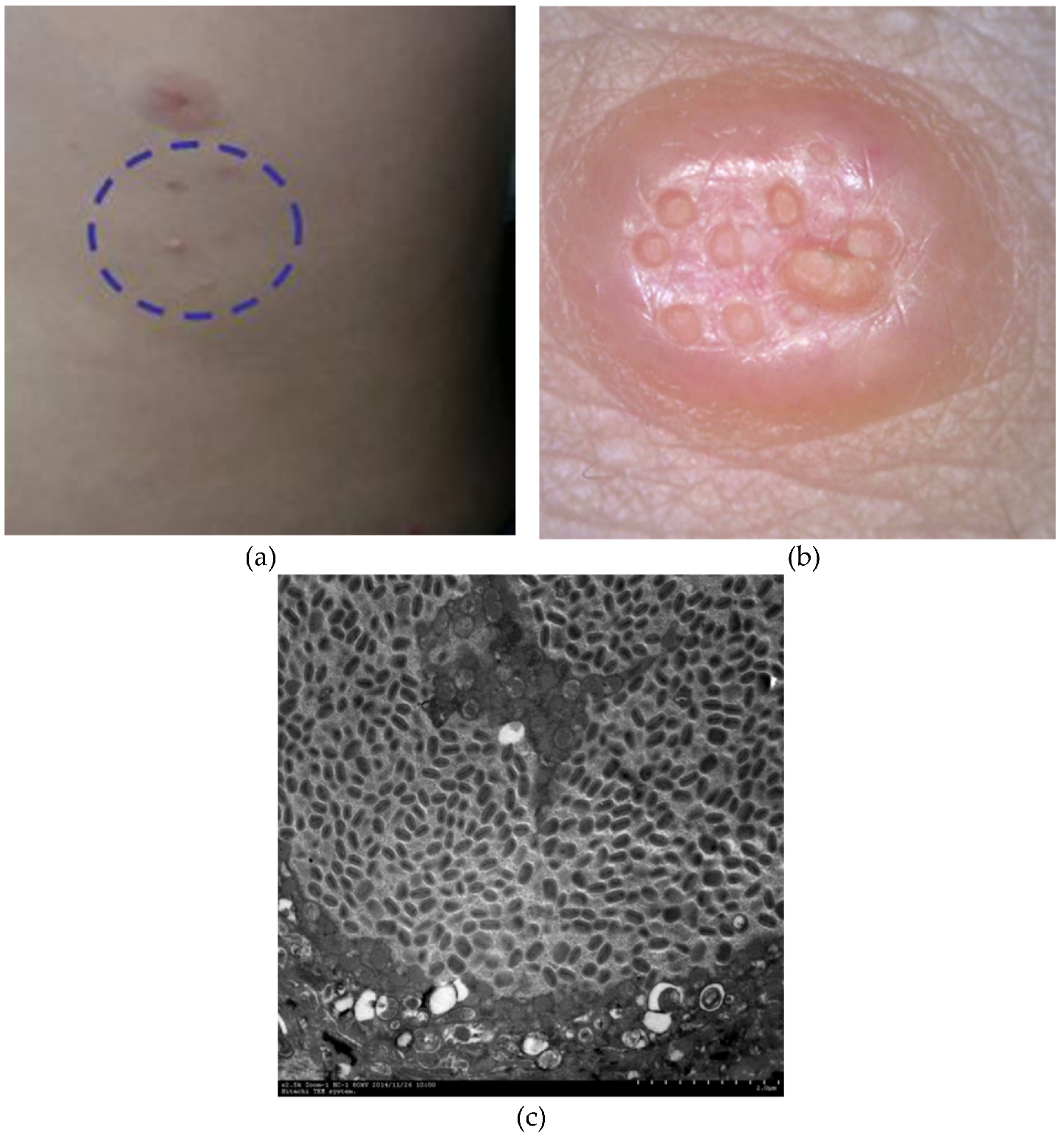
Molluscum contagiosum is a disease caused by a poxvirus. It is more prevalent in children up to 5 years of age. There is a second peak of incidence in young adults. In order to examine its ultrastructure, three lesions were curetted without disruption, cut transversely with a scalpel, and routinely processed for scanning electron microscopy (SEM). The oval structure of molluscum contagiosum could be easily identified. In its core, there was a central umbilication and just below this depression, there was a keratinized tunnel. Under higher magnification, a proliferation similar to the epidermis was seen. Moreover, there were areas of cells disposed like a mosaic. Under higher magnification, rounded structures measuring 0.4 micron could be observed at the end of the keratinized tunnel and on the surface of the lesion.
- [5]
Under lower magnification, the oval structure of MC could be easily identified. In its core, there was a central umbilication and just below this depression, there was a keratinized tunnel (
Figure 1). Under higher magnification, it was possible to observe the epidermis and the stratum corneum; underneath the epidermis, there was a proliferation similar to the epidermis (
Figure 2). Moreover, there were areas of cells disposed like a mosaic (
Figure 3).
- [5]
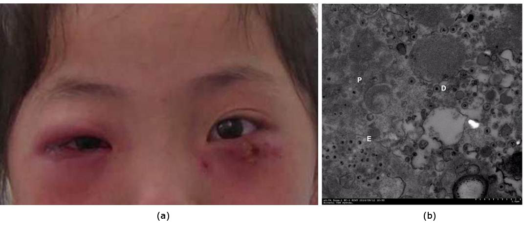
Herpes Simplex
Although capsids of herpes simplex virus were encountered within phagocytic vesicles, they were more commonly observed free within the cytoplasm. Stages in the release of virus from vesicles were not seen. There appeared to be five distinct steps in the process whereby the virus initiates infection: attachment, digestion of the viral envelope, digestion of the cell wall, passage of the capsid directly into the cytoplasm, and digestion of the capsid with release of the core. Antibody probably interferes with the first two stages.
- [6]
- https://www.ncbi.nlm.nih.gov/pmc/articles/PMC375640/?page=3
- https://www.ncbi.nlm.nih.gov/pmc/articles/PMC375640/?page=4
- https://www.ncbi.nlm.nih.gov/pmc/articles/PMC375640/?page=5
- https://www.ncbi.nlm.nih.gov/pmc/articles/PMC375640/?page=7
- https://www.ncbi.nlm.nih.gov/pmc/articles/PMC375640/?page=8
[3] Craig Freudenrich, Ph.D. "How Viruses Work" 19 October 2000.
[4] Relevant to Round 1 and found at the bottom of it.
[5] Almeida, H. L., Abuchaim, M. O., Schneide, M. A., Marques, L., & Castro, L. A. (2013). Scanning electron microscopy of molluscum contagiosum. Anais brasileiros de dermatologia, 88(1), 90-3.
[6] Morgan, C., Rose, H. M., & Mednis, B. (1968). Electron microscopy of herpes simplex virus. I. Entry. Journal of virology, 2(5), 507-16.

Not all earthlings are fools.
The idea that viruses don't exist is indefensible, it's like denying that the sun exists or that trees exist, we literally have viruses in laboratories right now.
That makes 7.4 billion and one. lol
7.4 billion
How many fools are there on planet Earth anyway?
Come on, this is not right...right?